CD169+ macrophages in immune regulationB6;129-Siglec1tm1(HBEGF)Mtka RBRC04395
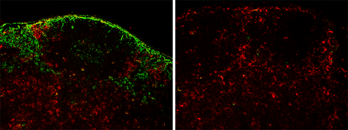
Selective depletion of CD169-positive cells in lymph node. |
Apoptotic cell death is a critical process for the elimination of unnecessary cells. After undergoing apoptosis, the remains of cells are phagocytosed by macrophages and dendritic cells. The rapid removal of apoptotic cells by phagocytes prevents the release of toxic and/or immunogenic materials. The injection of cell-associated exogenous antigens is known to induce tolerance to these antigens in mice. It is presumed that the injected apoptotic cells are phagocytosed by antigen-presenting cells (APCs) in the spleen, and that the APCs then present peptides derived from the apoptotic cells to T cells.
Dr. Tanaka and colleagues revealed that the injected apoptotic cells accumulate initially in the marginal zone (MZ) of the spleen. The role of macrophages in the MZ for tolerance induction was examined using human diphtheria toxin receptor (DTR) transgenic mice, in which macrophages expressing CD169 in the MZ are specifically ablated by means of the toxin receptor-mediated conditional cell knockout (known as TRECK) system [1]. Analysis of these mice revealed that macrophages in the MZ regulated both the clearance of circulating apoptotic cells and the selective engulfment of dying cells by CD8α+ dendritic cells. In the absence of these macrophages, the induction of tolerance to cell-associated antigens was severely impaired [2,3]. Recently, CD169+ macrophages have been identified as lymph node-resident APCs, dominating the early activation of tumor antigen-specific CD8+ T cells [4]. CD169-DTR mice provide the opportunity to clarify the role of CD169+macrophages in apoptotic cell clearance and T cell immunity.
| Depositor | : | Dr. Masato Tanaka School of Life Science, Tokyo University of Pharmacy and Life Sciences |
||||||||
| References | : |
|


