

| February 2017 Mouse of the Month |
Cell type-specific expression of photoconvertible fluorescent protein KikGR
B6.Cg-Gt(ROSA)26Sor<tm1(CAG-kikGR)Kgwa> (RBRC09254)
|
|
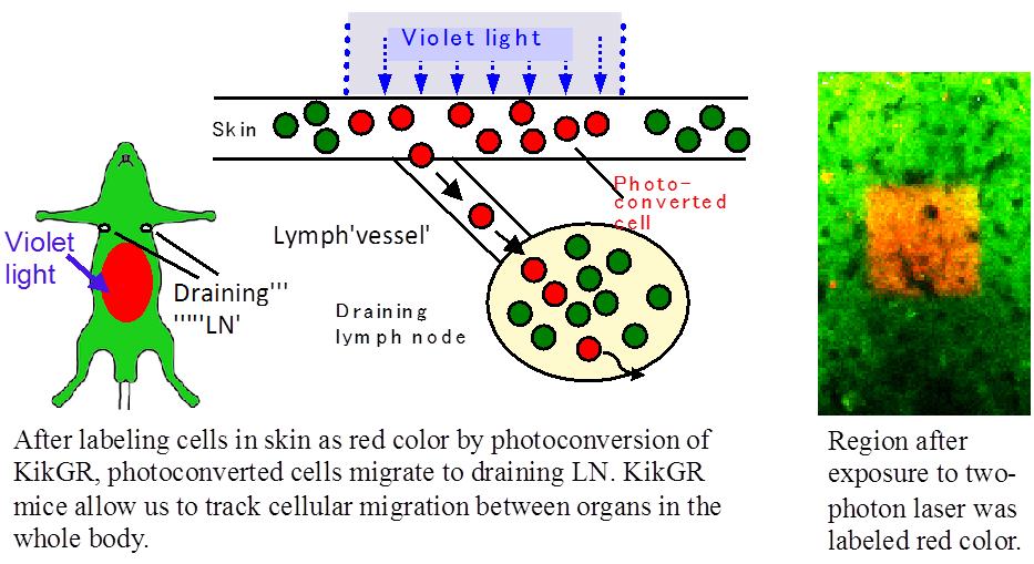
Kikume Green-Red protein (KikGR) is a photoconvertible fluorescent protein which was originally engineered from a stony coral Favia favus (“kikume-ishi” in Japanese) [1]. KikGR changes its color from green to red after exposure to violet light, and it has greater conversion efficiency compared to Kaede. Tomura and colleagues inserted CAG-LSL-KikGR DNA construct into ROSA26 locus and generated KikGR knock-in mouse strain, from which cell type-specific KikGR expression can be achieved by mating with Cre driver mice [2]. They labeled and tracked the migration and turnover of skin-derived dendritic cells [2], RANK+ splenocytes including osteoclast precursors [3], and tumor-infiltrating monocytes derived from bone marrow (BM) and spleen by using BM chimera mice [4]. KikGR knock-in mice are a new tool to monitor cell replacement at the photoconverted site and/or cell migration to other regions in the body. |
| Depositor | : | Haruhiko Koseki, M.D., Ph.D. Laboratory for Developmental Genetics RIKEN Center for Integrative Medical Sciences (IMS) |
|
| Strain name | : | B6.Cg-Gt(ROSA)26Sor<tm1(CAG-kikGR)Kgwa> | |
| RBRC No. | : | RBRC09254 | |
| Strain name | : | C.Cg-Gt(ROSA)26Sor<tm1(CAG-kikGR)Kgwa> | |
| RBRC No. | : | RBRC09255 | |
| References | : | [1] | Tsutsui H, Karasawa S, Shimizu H, Nukina N, Miyawaki A. Semi-rational engineering of a coral fluorescent protein into an efficient highlighter. EMBO Rep.; 6(3):233-8, 2005. |
| [2] | Tomura M, Hata A, Matsuoka S, Shand FH, Nakanishi Y, Ikebuchi R, Ueha S, Tsutsui H, Inaba K, Matsushima K, Miyawaki A, Kabashima K, Watanabe T, Kanagawa O. Tracking and quantification of dendritic cell migration and antigen trafficking between the skin and lymph nodes. Sci Rep.; 4:6030, 2014. | ||
| [3] | Kotani M, Kikuta J, Klauschen F, Chino T, Kobayashi Y, Yasuda H, Tamai K, Miyawaki A, Kanagawa O, Tomura M, Ishii M. Systemic circulation and bone recruitment of osteoclast precursors tracked by using fluorescent imaging techniques. J Immunol.; 190(2):605-12, 2013. | ||
| [4] | Shand FH, Ueha S, Otsuji M, Koid SS, Shichino S, Tsukui T, Kosugi-Kanaya M, Abe J, Tomura M, Ziogas J, Matsushima K. Tracking of intertissue migration reveals the origins of tumor-infiltrating monocytes. Proc Natl Acad Sci U S A.; 111(21):7771-6, 2014. | ||
| February 2017 Contact: Shinya Ayabe, Ph.D. Experimental Animal Division, RIKEN BioResource Center All materials contained on this site may not be reproduced, distributed, displayed, published or broadcast without the prior permission of the owner of that content. |