Visualizing neuronal activityTg(aMHC-VSFP2.3)107Bsi RBRC04705
|
| Schematic figure of the cortical barrel field |
 |
 |
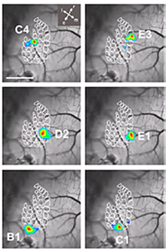
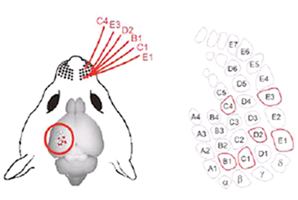
Figure 1. Sequential stimulation of single whiskers (C4, E3, D2, B1, C1, and E1) triggered electrical responses of neurons in the corresponding cortical area. Imaging of VSFP successfully detected neuronal activity in the corresponding cortical barrel (Nature Methods 7, 646, 2010).
The VSFP2 mouse is the most advanced optogenetic transgenic mouse strain generated using a newly developed voltage-sensitive fluorescent protein (VSFP2.3) and was deposited at RIKEN BRC by Dr. Thomas Knöpfel, RIKEN Brain Science Institute. By stimulating a single whisker of the VSFP2 transgenic mouse, the electrical responses of the pyramidal neurons in the corresponding somatosensory cortical barrel area can be visualized (Fig. 1). These optogenetic tools, which allow the visualization of neuronal electrical responses in real time, are expected to reveal neural circuit deficiencies in psychiatric disorder models.
| Depositor | : | Dr. Thomas Knöpfel, Imperial College London | |
| References | : | [1] | Lundby A, Mutoh H, Dimitrov D, Akemann W, Knöpfel T. Engineering of a genetically encodable fluorescent voltage sensor exploiting fast Ci-VSP voltage-sensing movements.PLoS One. 2008 3(6):e2514. |
| [2] | Akemann W, Mutoh H, Perron A, Rossier J, Knöpfel T. Imaging brain electric signals with genetically targeted voltage-sensitive fluorescent proteins. Nat Methods. 2010 7(8):643-9. | ||


